
Product overview
Foot Skeleton Model with Ligaments & Muscles

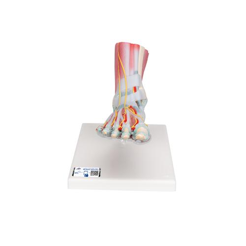
Foot Skeleton Model with Ligaments & Muscles is an anatomical teaching aid for examining the structures and features described below.
Product features and anatomical detail
- This anatomically detailed model of the foot and lower leg can be disassembled into 6 removable parts for detailed study of the foot and ankle.
- The foot skeleton features not only the bones but also the muscles, tendons, ligaments, nerves, arteries of the foot.
- The frontal view of the foot model features the extensor muscles of the lower leg.
- The tendons can be followed on their passage under the transverse and crucial crural ligaments all the way to their insertion points.
- In addition all tendon sheaths of the foot area are visible.
- On the dorsal portion of the foot the gastrocnemius muscle is removable to reveal deeper anatomical elements.
- The sole of the foot is represented in three layers.
- The first layer displaying the flexor digitorum brevis.
- This second layer is in turn removable to display even deeper anatomical details of the foot.
Product specifications
| Catalogue number | 1019421 [M34/1] |
|---|---|
| Weight | 1.2 kg |
| Dimensions | 23 x 26 x 19 cm |