

Ultrasound simulation · MU-E-BRE-MNS
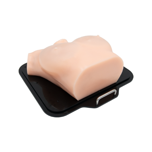
Breast Ultrasound Diagnosis Model (Skin Tone)
Product overview
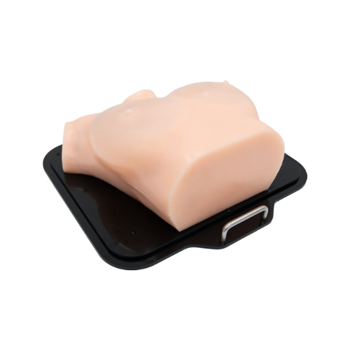
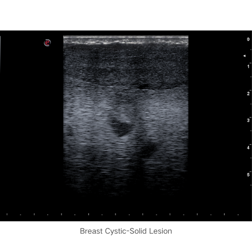
Practise breast ultrasound scanning, anatomical recognition, lesion detection and elastography using real ultrasound equipment. The adult female chest model represents skin, glands, ligaments and fat, with lesions of different echogenicity.
Training skills and applications
- Breast ultrasound anatomy demonstration
- Breast ultrasound examination technique training
- Training in size measurement, echo characteristics, location positioning, and shape description of common breast lesions
- Breast elastography ultrasound examination
Anatomy
- Female anatomy from neck to upper abdomen, including bilateral breasts, supraclavicular fossae, and axillae
- Palpable surface landmarks based on bony structures such as the manubrium sterni, bilateral clavicles, sternum, xiphoid process, and ribs
- Skin, fat, mammary glands, mammary ducts, lobules of the breast, Cooper's ligaments, subglandular fat, and pectoralis major muscles
Simulated pathology
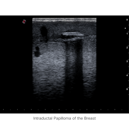
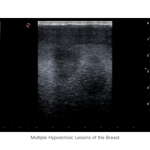
- Benign breast tumors, malignant breast tumors, breast cysts, mammary duct ectasia, mammary duct papillomas, axillary lymphadenopathy, supraclavicular fossa lymphadenopathy
Product features
Feature 1
Represents female breast shape and internal anatomical structures.
Feature 2
More than ten clinical cases vary in echogenicity and morphology.
Feature 3
Supports scanning throughout the breast quadrants and lymph-node regions.
Feature 4
Supports breast scanning technique, lesion recognition and elastography training.
Product specifications
- Catalogue number
- MU-E-BRE-MNS
- Colour
- Skin tone
- Pump
- None
Consumables — supplied separately
None
Related models and configurations
Product photographs
Downloads and supplier information
Plan your training
Contact Survival Technology to discuss this product and your ultrasound training requirements.