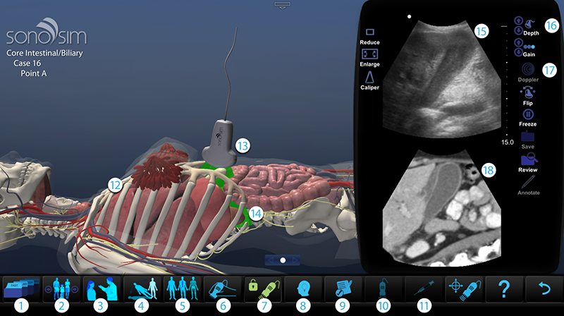
The SonoSimulator® provides the below basic feature set*.
The Case List button provides immediate access to a wide-ranging and ever-growing SonoSim® Case Library.
The Case Navigation feature allows users to navigate forward or backward to the next hands-on training case by selecting the arrow icons on the Case button.
The Case History feature provides a brief patient history including information such as age, gender, vital signs, and chief complaint.
The Body Position feature allows users to place the virtual patient in different positions that provide alternative viewer perspectives.
With the Layers feature, anatomic layers can be removed to reveal underlying anatomy, allowing the user to correlate the ultrasound image to relevant anatomic structures.*
Probe-positioning guidance assists users in orienting the probe in the optimal imaging plane.
Lock the probe in place to maintain the ultrasound window.
The Findings button provides immediate expert feedback. A narrated version of the ultrasound scan describes what users should recognize while scanning the corresponding SonoSim® Case.
Test Mode disables assistive features such as Findings, Probe Guide, and Layers in order to test learners’ mastery of image acquisition and interpretation.
Hold the Compression button to compress the structure immediately beneath the transducer (e.g., compress a vein).*
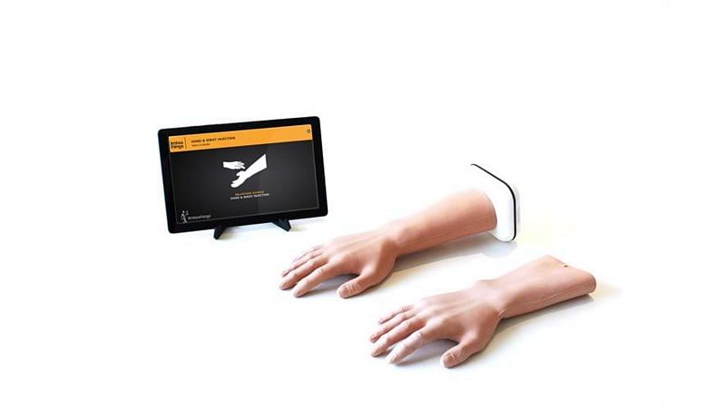
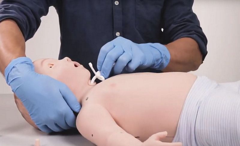
The Needle procedure feature provides cognitive task training on ultrasound-guided needle-based procedures.*
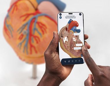
A three-dimensional human body model depicts dynamic anatomic structures. Adult & pediatric and male & female bodies are displayed as are appropriate.
This helps the user recognize which transducer to use when performing an actual scan.
Learn how the array beam traverses anatomical structures to create the corresponding ultrasound image in the display window.
Ultrasound display window depicts images that correspond to probe movements with extremely high fidelity and real-time performance.
The ultrasound display window simulates a real ultrasound unit, including the ability to modify image depth and gain; flip the probe indicator; freeze, save, and review obtained images; and measure anatomic structures.
The Doppler button enables learners to examine motion detection and blood flow characteristics through a variety of imaging modalities, including m-mode and power, color-flow, and pulsed-wave Doppler.*
The Split Screen feature enables users to easily compare ultrasound findings and image characteristics with those provided by other imaging modalities (e.g., plain radiographs, CT, or MRI).*
*These advanced features are only included in specific SonoSim® Modules.
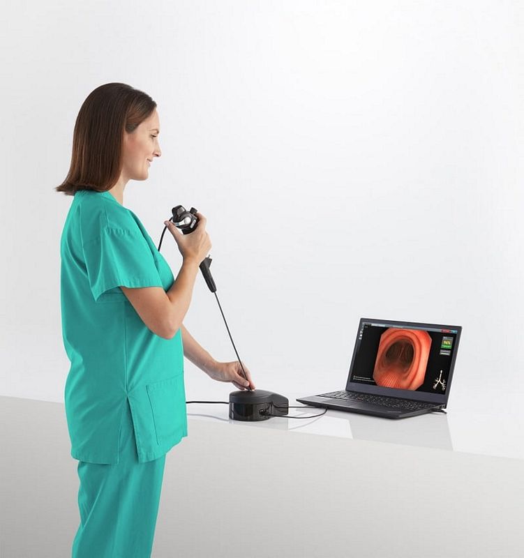
Experience SonoSimulation®, the process through which real patient ultrasound data is converted into laptop-based, personalized ultrasound training solution.

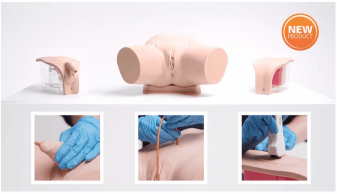
The SonoSim® Ultrasound Training Solution can be configured to provide cognitive task training on a variety of ultrasound-guided needle-based procedures. This allows you to develop hand-eye coordination in a safe environment. You are provided unlimited practice opportunities through the use of real patient-based virtual models.
The Case List button provides immediate access to a wide-ranging and ever-growing SonoSim® Case Library.
Users learn how to move an ultrasound transducer and localize a target vessel in short-axis and long-axis views.
Trainees learn how to achieve optimal needle penetration of a target vessel under ultrasound guidance. They learn the critical task of how to localize the tip of the needle by fanning the virtual ultrasound probe in an appropriate direction.
Probe-positioning assistance, for Longitudinal and Transverse window acquisition, allows for virtual “hand-holding” during image acquisition.
Users are given specific, real-time feedback following each ultrasound-guided intravenous catheter insertion attempt.
.png)


.png)